Description
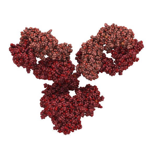
MOUSE ANTI-DENGUE PAN-SEROTYPE ENVELOPE PROTEIN ANTIBODY
Dengue pan-serotype envelope antibody reacts with envelope protein from Dengue virus serotypes 1, 2, 3 and 4. Antibody does not recognise DENV envelope protein domain III (EDIII).
PRODUCT DETAILS – MOUSE ANTI-DENGUE PAN-SEROTYPE ENVELOPE PROTEIN ANTIBODY
- Mouse anti-Dengue pan serotype Envelope monoclonal IgG2a antibody (clone M769).
- Greater than 95% purity by SDS-PAGE and buffered in 0.015 M potassium phosphate buffer, 0.85% NaCl, pH 7.2.
BACKGROUND
Dengue virus is a member of the flavivirus family, which includes Zika virus, West Nile virus and Japanese Encephalitis virus. Many of the proteins expressed by these viruses are very similar, and serological testing for these viruses can be complicated by problems of cross-reactivity. In this context, the use of highly purified antigens, that are glycosylated and folded as native proteins can be highly important in the development of accurate immunoassays.
With more than one-third of the world’s population living in areas at risk of transmission, dengue infection is a leading cause of illness and death in the tropics and subtropics (ref WHO for Guidelines for Diagnosis, Treatment, Prevention & Control, 2009). Dengue is caused by any one of four related viruses (DENV1-4) which are transmitted by mosquitoes and responsible for infecting as many as 390 million people yearly (Bhatt , et al., 2013).
It is a febrile illness that affects infants, young children and adults with symptoms appearing 3-14 days after the infective bite. Symptoms range from mild fever, to incapacitating high fever, with severe headache, pain behind the eyes, muscle and joint pain, and rash. There is no vaccine or any specific medicine to treat dengue.
Severe dengue (also known as dengue hemorrhagic fever) is characterized by fever, abdominal pain, persistent vomiting, bleeding and breathing difficulty and is a potentially lethal complication, affecting mainly children. Early clinical diagnosis and careful clinical management by trained physicians and nurses increase survival of patient.
More recently there has been increased interest in the potential of antibody-based immunotherapy as treatment for dengue. The dengue envelope (E) protein is arranged as antiparallel homodimers on the surface of the mature virion, comprising a total of 180 E proteins on the virion surface (Zhang, et al., 2013). It is composed of three domains, DI-DIII, in which DII contains the fusion loop required for viral entry (Modis, et al., 2004). An engineered cross-neutralizing humanized antibody targeting the DENV E domain III has been demonstrated to neutralize DENV in mice, at clinically relevant viral loads or in the presence of enhancing levels of DENV immune sera (Budigi, et al., 2018).
REFERENCES
- Bhatt , S. et al., 2013. The global distribution and burden of dengue. Nature, 496(7446), pp. 504-7.
- Budigi, Y. et al., 2018. Neutralization of antibody-enhanced dengue infection by VIS513, a pan serotype reactive monoclonal antibody targeting domain III of the dengue E protein. PLoS Negl Trop Dis., 12(2).
- Modis, Y., Ogata, S., Clements, D. & Harrison, S. C., 2004. Structure of the dengue virus envelope protein after membrane fusion. Nature, Volume 427, pp. 313-319.
- Zhang, X. et al., 2013. Cryo-EM structure of the mature dengue virus at 3.5-A resolution. Nat Struct Mol Biol , Volume 20, pp. 105-110.


