Description
MOUSE ANTI-ENTEROVIRUS 71 VP1 (3656)
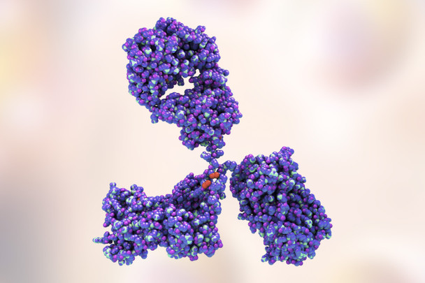
Mouse anti Enterovirus 71 VP1 (3656) antibody is specific for EV71 VP1 and has been developed for use in ELISA and immunofluorescence. These monoclonal antibodies have been developed to meet the need for highly reactive and specific EV71 antibodies for the future development of serological assays.
PRODUCT DETAILS – MOUSE ANTI-ENTEROVIRUS 71 VP1 ANTIBODY (3656)
- Mouse anti Enterovirus 71 VP1 (3656). Specific for the VP1 of Enterovirus type 71. Does not cross react with related viruses including Coxsackie A9, Coxsackie B2, ECHO 30, EV70 and Polio, type 2.
- Purified preparations consist of >90% pure mouse monoclonal antibody purified from ascites fluid or culture medium by protein A chromatography or sequential differential precipitations.
- Presented in PBS pH7.2 with 0.1% sodium azide.
- For use in ELISA and immunofluorescence.
- Can be used with Goat anti mouse IgG HRP and PanBlock ELISA Blocking Buffer.
BACKGROUND
Enteroviruses (EV) are single-stranded RNA viruses belonging to the Picornaviridae family and are the smallest, non-enveloped viruses known to infect both humans and animals. They are common seasonal viruses that are associated with a variety of diseases. Enterovirus 71 (EV71) has been identified as one of the main causative agents responsible for large outbreaks of hand, foot, and mouth disease (HFMD) across the Asia-Pacific region. Other members of the genus Enterovirus cause HFMD, including coxsackieviruses A16, A6, A5, A7, A9, A10, B2, and B5, but EV71 is linked to severe complications, including brainstem encephalitis, aseptic meningitis, and pulmonary edema (Caine et al., 2016). As yet, no effective EV-specific antiviral treatments are available, and vaccines are available only against polioviruses. Ongoing experience with EV71 outbreaks in the Asia-Pacific region has demonstrated that co-infections with other EV and indeed viruses belonging to other families, is common and raises the possibility that some co-infections can increase the severity of disease and change the clinical presentation.
EV are non-enveloped, and the virions are relatively simple, consisting of a protein capsid surrounding a single-stranded, positive sense RNA genome. The genome has approximately 7500 nucleotides, and contains a single open reading frame that encodes a polyprotein which is then processed to yield the structural (i.e., capsid) proteins VP1, VP2, VP3, and VP4 and the non-structural proteins. Once viral RNA has entered the host cell cytoplasm it is translated into a single polyprotein, although a second open reading frame (ORF) has been observed in some enterovirus genomes. The polyprotein is then cleaved by the viral proteases 2A and 3C into 10 proteins including capsid proteins and other replication proteins such as the RNA-dependent RNA polymerase 3D (3Dpol). 3Dpol initiates the synthesis of the negative-stranded copy of the genome making a dsRNA intermediate, which becomes the template to generate new positive-stranded genomes. Replication of viral RNA occurs in replication organelles that derive from host membranes which are induced upon viral infection. The newly synthesized positive-stranded genome is packaged into virions for release out of the cell. The virions are assembled into protomers and pentamers using the capsid proteins VP0, VP1 and VP3. VP1, VP2, and VP3 are exposed on the capsid surface, while VP4 is present inside the capsid. After the RNA is packaged into the virion, VP0 is processed into VP2 and VP4, which results in mature enterovirus virions. The enteroviruses then exit the cell through cell lysis to infect neighbouring cells although non-lytic pathways may also be used.
The VP1 capsid gene is considered to be an ideal target for typing enterovirus because of variability of the region. Phylogenetic analysis of VP1 gene sequences also correlates well with serotyping in serum neutralization assays (Muehlenbachs et al., 2015). A domain has also been identified on the EV71 VP1 capsid protein which is critical for receptor attachment and necessary for the stability of infectious virions. It includes a group of positively charged amino acids near the 5-fold vertices of the capsid protein. EV71 receptors are believed to increase the ability of the virus to become neurotropic and so increase the severity of the disease it causes (Caine et al., 2016). This region is therefore an ideal target for the development of neutralising antibodies and anti-viral drugs.
REFERENCES
- Enterovirus surveillance guidelines. Guidelines for enterovirus surveillance in support of the Polio Eradication Initiative. World Health Organization 2015.
- Jia et al. (2017). Effective in vivo therapeutic IgG antibody against VP3 of enterovirus 71 with receptor-competing activity. Sci Rep. 7:46402.
- Wells, AI and Coyne, CB (2019). Enteroviruses: A Gut-Wrenching Game of Entry, Detection, and Evasion. Viruses 2019, 11(5), 460.
- Caine et al. (2016). A Single Mutation in the VP1 of Enterovirus 71 Is Responsible for Increased Virulence and Neurotropism in Adult Interferon-Deficient Mice. J Virol. 90(19):8592-604.
- Muehlenbachs et al. (2015). Tissue tropism, pathology and pathogenesis of enterovirus infection. J Pathol. 235(2):217-28.
- Factsheet about enteroviruses. European Centre for Disease Prevention and Control (ECDC), 2010.


