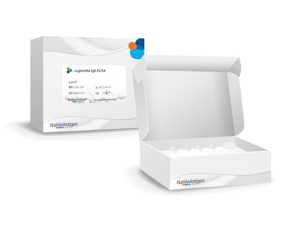
Description
LEGIONELLA PNEUMOPHILA IgM ELISA
Legionella pneumophila IgM ELISA for the qualitative determination of IgM class antibodies against Legionella pneumophila in human serum or plasma (citrate, heparin).
The qualitative immunoenzymatic determination of specific antibodies is based on the ELISA (Enzyme-linked Immunosorbent Assay) technique. Microplates are coated with specific antigens to bind corresponding antibodies of the sample. After washing the wells to remove all unbound sample material a horseradish peroxidase (HRP) labelled conjugate is added. This conjugate binds to the captured antibodies. In a second washing step unbound conjugate is removed. The immune complex formed by the bound conjugate is visualized by adding Tetramethylbenzidine (TMB) substrate which gives a blue reaction product. The intensity of this product is proportional to the amount of specific antibodies in the sample. Sulphuric acid is added to stop the reaction. This produces a yellow endpoint colour. Absorbance at 450/620 nm is read using an ELISA microwell plate reader.
Serologic testing for L. pneumophila serogroups is often the primary method of screening for possible L. pneumophila infections. Therefore, high sensitivity is paramount in a screening assay, since the assay should detect the greatest possible number of samples positive for L. pneumophila antibodies. The sensistivity of this assay is 100%.
PRODUCT DETAILS – LEGIONELLA PNEUMOPHILA IgM ELISA
-
- Legionella pneumophila IgM ELISA.
- High sensitivity – 100%.
- High specificity – 95.65%.
- Short assay time – <3 hours.
- 1 x 96 tests.
BACKGROUND
Legionella pneumophila is a thin, pleomorphic, flagellated Gram-negative bacterium of the genus Legionella and is the causative agent of legionellosis or Legionnaires’ disease. They are found in freshwater environments worldwide and can cause respiratory disease (legionellosis) in humans. Strains of the organism were first isolated in the 1940s but only came to prominence after an outbreak of pneumonia involving delegates of the 1976 American Legion Convention at a Philadelphia hotel. The genus Legionella currently has at least 50 species comprising 70 distinct serogroups. One species of Legionella, L. pneumophila, is the aetiological agent of approximately 90 % of legionellosis cases, and serogroup 1 (Sg1) accounts for about 84 % of these cases (WHO).
L. pneumophila multiplies itself at temperatures between 25 and 42 °C, with an optimal growth temperature of 35 °C. Legionella thrives in warm, stagnant water in the environment and in artificial systems such as cooling towers, evaporative condensers, hot and cold-water systems and spa pools that mimic the natural environment in which the organism thrives. These systems also provide the means by which aerosols/droplets are generated and the organism dispersed into the atmosphere. The most common form of transmission of Legionella is inhalation of contaminated aerosols produced in conjunction with water sprays, jets or mists. Infection can also occur by aspiration of contaminated water or ice, particularly in susceptible hospital patients. Person-to-person transmission is not thought to be a risk. In Europe, Australia and the USA there are about 10–15 cases detected per million population per year and there is no vaccine currently available for Legionnaires’ disease.
The likelihood of contracting Legionnaires’ disease depends on the level of contamination in the water source, the susceptibility of the person exposed, and the intensity of exposure. Legionnaires’ disease is characterized as an “opportunistic” disease that attacks individuals who have an underlying illness or a weakened immune system. Predisposing risks include increasing age, being male, heavy smoking, alcohol abuse, chronic lung disease, immunosuppressive therapy, cancer chemotherapy, organ or bone marrow transplant, and corticosteroid therapy. Legionellosis can appear in two distinct clinical presentations: Legionella pneumonia (Legionnaires’ disease) with an incubation period of approx. 2-10 days (may extend up to 16-20 days) and Pontiac fever (incubation period: normally 12-48 hours). Legionella pneumonia (Legionnaires’ disease) is a serious form of pneumonia that carries with it a case-fatality ratio of 10-15 %. Legionnaires’ disease patients initially present with cough, fever and nonspecific symptoms including malaise, myalgia and headache. Some patients develop shaking chills, chest pain, diarrhea, delirium or other neurologic symptoms. Extra pulmonary involvement is rare.
Pontiac fever is a milder form of the disease without manifestations of pneumonia and presents as an influenza-like illness. Symptoms may include headache, chills, muscle aches, a dry cough and fever. It is usually self-limiting and typically does not require treatment. The attack rate is much higher than for Legionnaires’ disease (up to 95 % of those exposed).
REFERENCES
- Bartram, Jamie; Chartier, Yves; Lee, John V.; Pond, Kathy; Surman-Lee, Susanne (Eds.) (2007): Legionella and the prevention of legionellosis. Geneva: WHO (14).
- Darby, Jonathan; Buising, Kirsty (2008): Could it be Legionella? In Australian Family Physician 37 (10), pp. 812–815.
- Fields, Barry S.; Benson, Robert F.; Besser, Richard E. (2002): Legionella and Legionnaires’ Disease. 25 Years of Investigation. In Clinical Microbiology Reviews 15 (3), pp. 506–526. DOI: 10.1128/CMR.15.3.506-526.2002.
- Joseph, C. A. (2004): Legionnaires’ disease in Europe 2000-2002. In Epidemiology and infection 132 (3), pp. 417–424. DOI: 10.1017/S0950268804002018.
- Marrie, Thomas J.; Hoffman, Paul (2006): Legionellosis. In Richard L. Guerrant, David H. Walker, Peter F. Weller (Eds.): Tropical infectious diseases. Principles, pathogens & practice. 2nd ed. Philadelphia: Churchill Livingstone, pp. 374–380.
- Robert Koch Institut (RKI) (2013): Legionellose. In RKI-Ratgeber für Ärzte.
- Steinert, Michael; Hentschel, Ute; Hacker, Jörg (2002): Legionella pneumophila: an aquatic microbe goes astray. In FEMS microbiology reviews 26 (2), pp. 149–162.
- Stout, Janet E.; Yu, Victor L. (1997): Legionellosis. In The New England Journal of Medicine 337 (10), pp. 682–687. DOI: 10.1056/NEJM199709043371006.
- Yu, Victor L.; Plouffe, Joseph F.; Pastoris, Maddalena Castellani; Stout, Janet E.; Schousboe, Mona; Widmer, Andreas et al. (2002): Distribution of Legionella species and serogroups isolated by culture in patients with sporadic community-acquired legionellosis: an international collaborative survey. In The Journal of Infectious Diseases 186 (1), pp. 127–128. DOI: 10.1086/341087.
- Zuravleff, Jeffrey J.; Yu, Victor L.; Shonnard, John W.; Davis, Bridgett K.; Rihs, John D. (1983): Diagnosis of Legionnaires’ disease. An update of laboratory methods with new emphasis on isolation by culture. In JAMA 250 (15), pp. 1981–1985.
THIS ELISA ASSAY IS FOR RESEARCH USE ONLY. IT IS NOT FOR USE IN DIAGNOSTIC PROCEDURES.


